Metastasis is not driven by a uniform population of cancer cells. Tumour cells can change their phenotype in response to intrinsic and microenvironmental signals, generating states with different capacities to migrate, survive, interact with host cells and respond to treatment. We investigate this plasticity across the metastatic cascade, from the primary tumour to cells detected in the circulation and metastatic sites.


Research overwiew
Breast cancer is our principal disease model. A central theme of our work is epithelial–mesenchymal transition (EMT), a dynamic programme associated with changes in adhesion, motility, cellular identity and tumour-cell behaviour.
Epithelial–mesenchymal plasticity

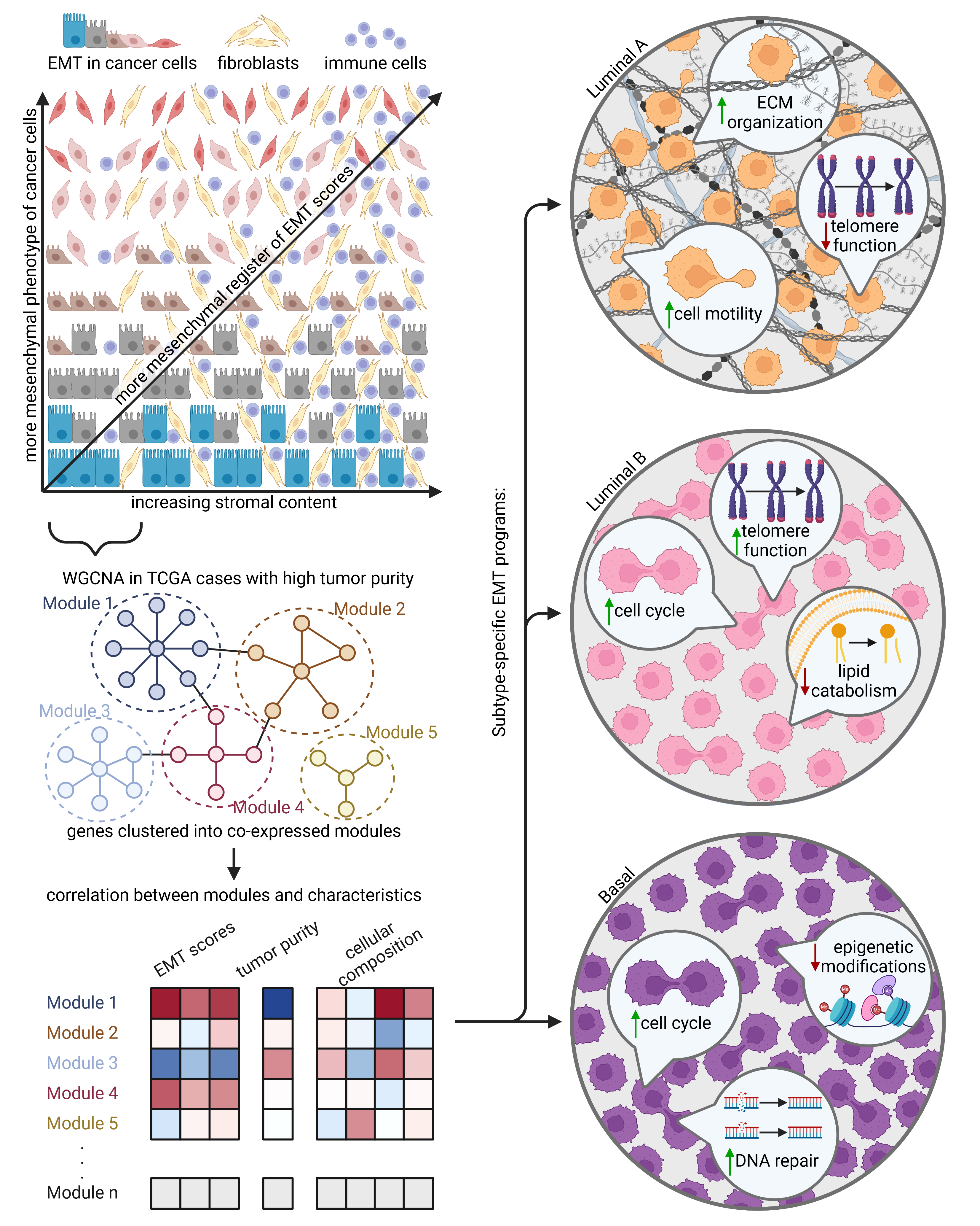
Epithelial–mesenchymal transition is a major source of phenotypic diversity in cancer. Our work examines what EMT-associated states arise during breast cancer progression and how they are represented in primary tumours, lymph-node metastases and circulating tumour cells. We are particularly interested in the relationship between EMT state and the aggressive properties required for successful dissemination of cancer cells at different sites of the body. Rather than treating EMT as a binary switch and linear sequence of changes, we investigate the different trajectories of EMT phenotypes depending on intrinsic molecular profile of cancer cells.
Our research has shown that breast cancer encompass a broad spectrum of EMT phenotypes and that this heterogeneity. We use this framework to ask which molecular programs accompany distinct EMT states and which features may create vulnerabilities that can be exploited therapeutically. If EMT drives aggressive behaviour of cancer cells – can it be blocked or reversed in order to limit cancerous growth or individual properties, which make cancer a deadly disease?
Liquid biopsy approaches in tracking cancer dissemination
Solid tumours might shed their cells, vesicles or nucleic acids to circulation, which gives a unique opportunity to sample these oncogenic signals straight from blood or other body fluids (so called Liquid biopsy concept). In such way signals origination from cancer cells can be captured repeatedly in fairly non-invasive way (drawing a sample of venous blood), allowing for longitudinal study of tumour evolution and dissemination even beyond the primary lesion.
Circulating tumour cells
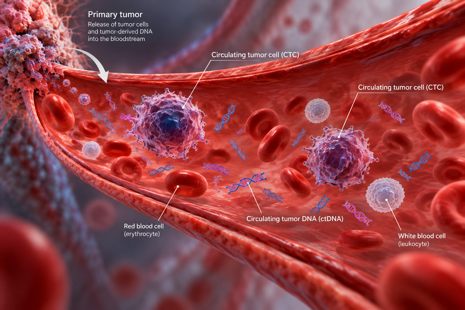
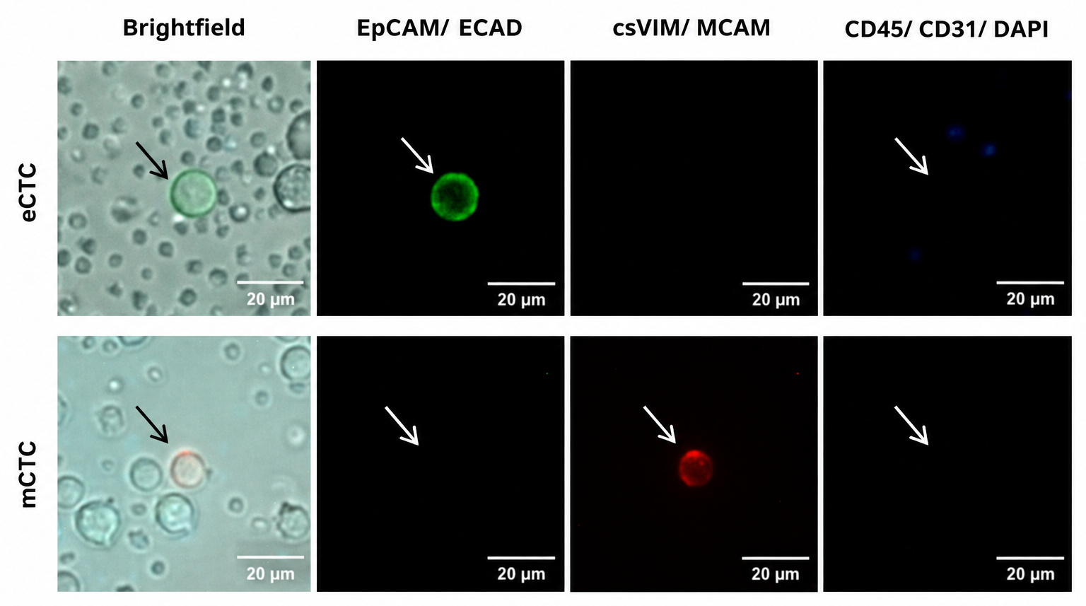
Circulating tumour cells (CTCs) are rare cells shed from tumour lesions into the circulation or other body fluids and represent a direct cellular component of the metastatic process. Our team applies approaches that recover and characterize CTCs with different phenotypes, including cells that may be missed by methods relying exclusively on epithelial markers. We combine phenotypic identification with downstream single-cell genomic (mutation, genomic aberrations assessed via a number of methods – MassArray, seqencing) and transcriptomic analysis (RNA-Seq, qPCR) to investigate the molecular diversity of individual CTCs and their relationship to metastatic progression.
Circulating tumour DNA
Cancer cells might actively or passively (due to cell death) release their DNA to circulation, where it can be detected and thus informing about genotypes of cancer clones present in the body and show how cancer evolvs. This also allows for monitoring of minimal residual disease (MRD) – detecting traces of tumour even if tumour leasions themselves are too small to be seen by imaging methods.
Computational analysis
We are using advanced statistical analysis and computational tools to analyze bulk and single-cell RNA-Seq profiles, clinico-pathological data to deciphering biological processes occurring in tumours and CTCs, what drives further hypothesis and experimental research.