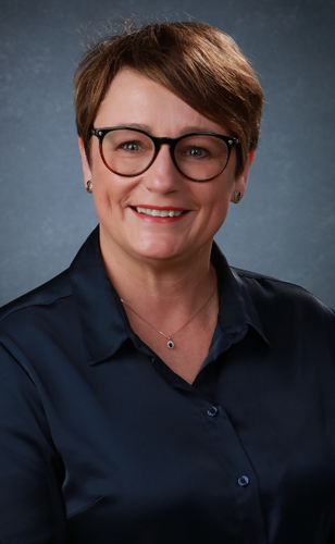
Wykład otwarty prof. Iwony Dobruckiej

Zakład Medycznej Diagnostyki Laboratoryjnej jest organizatorem spotkania z prof. Iwoną Dobrucką z University of Illinois, która wygłosi w jego trakcie wykład otwarty pt. Zastosowanie technik obrazowych PET (pozytronowa tomografia emisyjna) w różnych modelach zwierzęcych. Spotkanie odbędzie się 27 listopada 2023 r. w godz. 14:10-16:10 w auli im. prof. Zdzisława Kieturakisa w budynku Centrum Medycyny Inwazyjnej. Przeznaczone jest ono zarówno dla studentów, jak i pracowników GUMed
Zapisy dla studentów odbywają się za pośrednictwem strony szkoleniazawodowe.gumed.edu.pl.
PRELEGENTKA
Profesor Iwona Dobrucka dołączyła do Zakładu Bioinżynierii w marcu 2020 r. Badaczka uzyskała doktorat z chemii na Uniwersytecie Ohio w Athens, Ohio w 2004 r. oraz tytuł magistra bioinżynierii na Uniwersytecie Technicznym we Wrocławiu i Uniwersytecie Technicznym w Hamburgu w Niemczech. Przed dołączeniem do Beckman Institute w 2011 r., gdzie kieruje Laboratorium Obrazowania Molekularnego (MIL) w Biomedical Imaging Center (BIC), specjalistka szkoliła się z biologii raka w Katedrze Terapii Promacyjnej w Yale University School of Medicine w New Haven, Connecticut. Do jej zainteresowań należą takie obszary badawcze jak:
- obrazowanie molekularne
- molekularny PET-CT
- sondy bioobrazowe nanocząsteczkowe
- nanosensory
- diagnostyka optyczna w przypadku raka.
Obecnie skupia się na badaniu interakcji między tradycyjnymi lekami przeciwnowotworowymi a uzupełniającymi lekami alternatywnymi (CAM) stosowanymi przez pacjentów chorych na raka.
Iwona Teresa Dobrucka Ph. D.Ph.D. in chemistry, M.Sc. in biomedical engineering, and postdoctoral training in imaging and therapeutic radiology. Extensive (>10 years) working knowledge of small animal microSPECT-CT, microPET-CT hybrid and dedicated microCT imaging systems. Experienced with optical imaging, ultrasonography, microPET-CT imaging, and the design of implantable biosensors to detect reactive oxygen and nitrogen species in vivo. Currently directing the Molecular Imaging Laboratory within the Biomedical Imaging Center at Beckman Institute. Molecular Imaging Laboratory (MIL) is equipped with state-of-the-art hybrid microPET/SPECT/CT and 3D-optical-CT scanners and a complete molecular/cell biology and radiochemistry resource. Possesses the required expertise, experience, and motivation to lead and significantly contribute to the proposed research program, including radiolabeling and quality control of imaging agents, and performing both surgical procedures and animal imaging studies. She has a broad background in instrumental analysis, radiochemistry, animal physiology, microsurgery, and translational bioengineering, with specific training and expertise in key research areas for this application.
Since her postdoctoral training in imaging and therapeutic radiology at Yale University, she has been actively involved in the design and implementation of advanced imaging strategies, including the development of uni- and multimodal agents, validation in vitro and in vivo studies, and implementation of advanced imaging protocols to non-invasively assess biological processes in animal models of disease including peripheral and myocardial angiogenesis, arteriogenesis, atherosclerosis, vascular remodeling, cerebral stroke, and cancer. Her current research is focused on studying interactions between traditional anti-cancer therapeutics and complementary alternative medicines (CAMs) used by cancer patients.
